What Actually Happens During Autophagy
The word autophagy translates from Greek as "self-eating" — autos (self) and phagomai (to eat). The name sounds dramatic, and the process it describes is not far off. Your cells literally consume their own damaged parts, breaking them down into raw materials and reassembling those materials into functional components. Think of it as a factory that melts down its own broken machinery, recasts the metal, and builds new equipment from it.
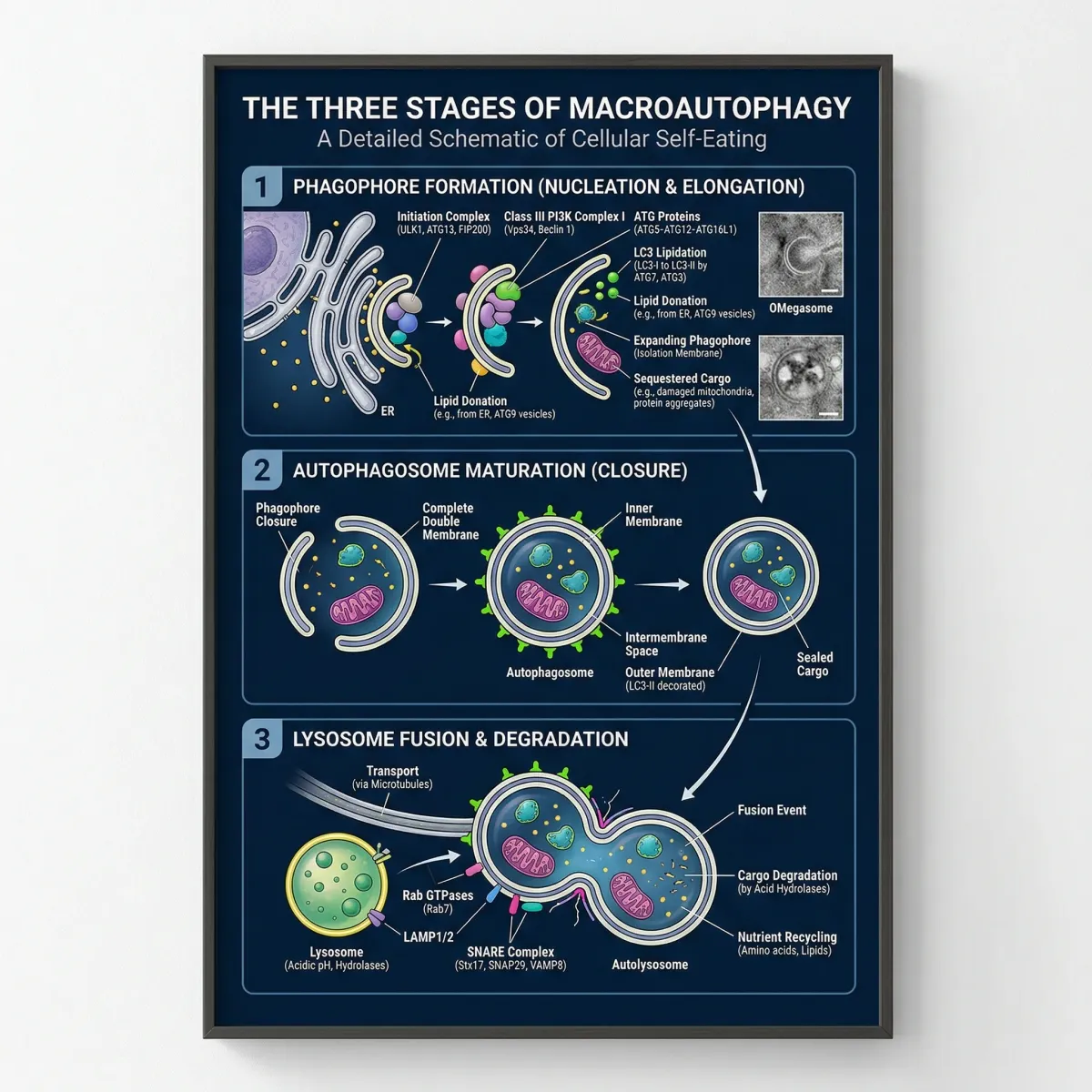
There are three recognized forms of this process. Macroautophagy is the primary type and the one most research focuses on. It wraps damaged organelles and protein aggregates inside double-membraned structures called autophagosomes, then delivers those packages to lysosomes — the cell's digestive compartments, for breakdown. Microautophagy skips the autophagosome step entirely; lysosomes directly engulf small bits of cytoplasmic material through their own membrane. Chaperone-mediated autophagy is the most selective of the three: specific heat shock proteins recognize a particular amino acid sequence on target proteins and shuttle them directly into lysosomes.
The molecular machinery driving macroautophagy involves a family of autophagy-related proteins (ATGs). These proteins orchestrate the formation of the phagophore (the initial membrane cup), its elongation into a sealed autophagosome, and its eventual fusion with a lysosome. Key players include ULK1 kinase, which initiates the process, and LC3 protein, which gets incorporated into the autophagosome membrane and serves as the most commonly measured marker of autophagy activity in laboratory studies.
Why Your Cells Need This Built-In Cleanup System
Cells accumulate damage constantly. Metabolic reactions generate reactive oxygen species that oxidize proteins. Mitochondria wear out and start leaking electrons. Misfolded proteins clump together into aggregates that gum up normal cellular operations. Without a reliable disposal system, this junk builds up, and the consequences show up as disease.
Autophagy handles what your cells cannot delegate to anyone else. It removes protein aggregates linked to neurodegenerative conditions like Alzheimer's and Parkinson's disease. It clears out dysfunctional mitochondria through a specialized process called mitophagy (requiring PINK1 and Parkin receptors). It even destroys intracellular pathogens including certain viruses and bacteria, acting as an immune defense layer that operates below the threshold of your broader immune system.
Key fact: Animal studies have shown that mice with disabled autophagy genes age faster and die earlier than normal mice. Conversely, elevating autophagy activity extends lifespan in the same species.
The decline matters. As you age, autophagy efficiency drops. The Cleveland Clinic notes that this age-related decrease "can lead to a build-up of cellular junk parts." That accumulation correlates with diseases that cluster in later life: heart disease, kidney disease, liver disease, type 2 diabetes, and the neurodegenerative conditions already mentioned. Research published in Frontiers in Psychology frames it bluntly — the absence of autophagy in the elderly is one of the main causes of biological "waste" accumulation reflected in damaged proteins and organelles.
This is what makes the three natural triggers — fasting, exercise, and certain dietary compounds — worth examining closely. They each activate autophagy through overlapping but distinct molecular pathways, and the research on each has matured enough to draw practical conclusions.
Fasting as an Autophagy Switch
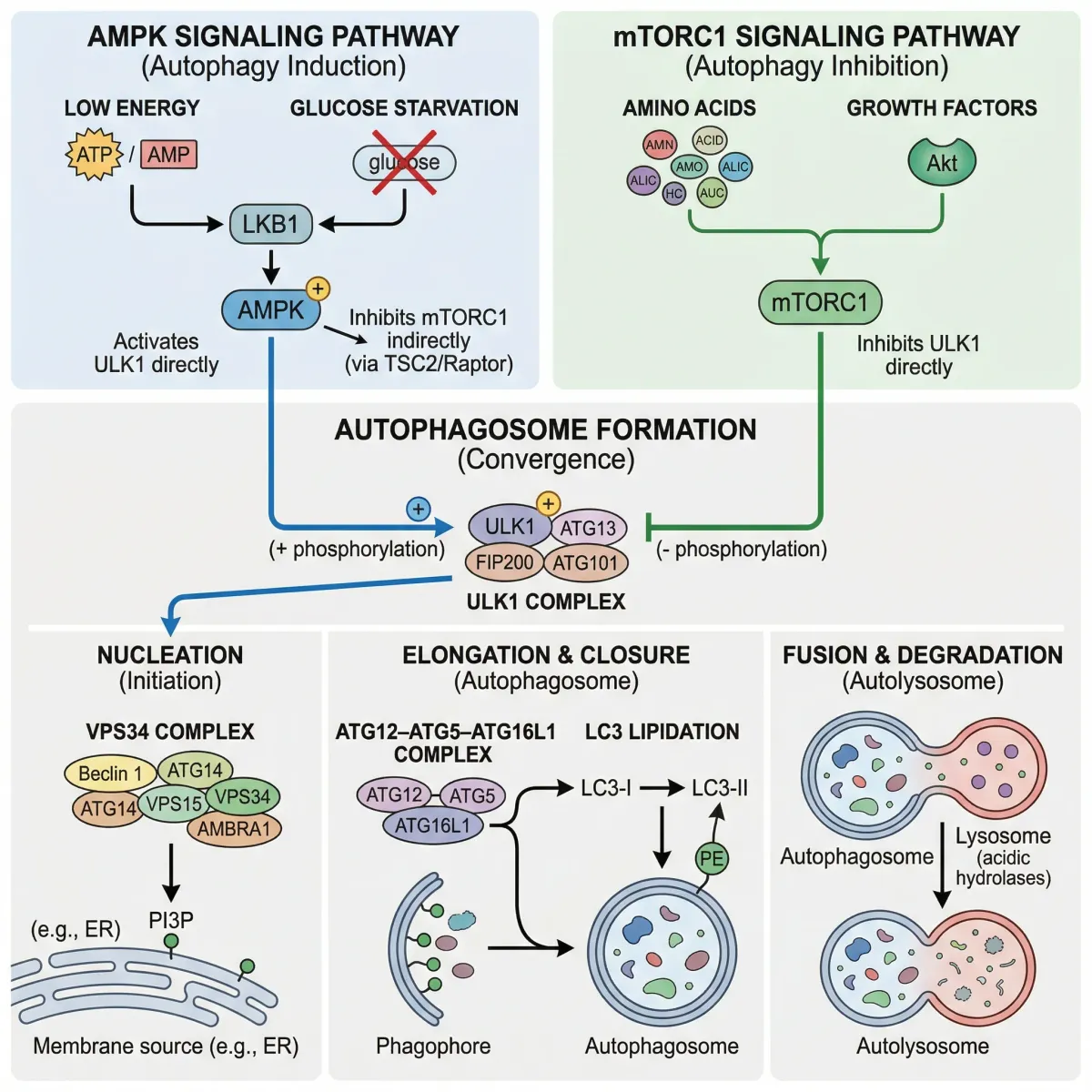
Fasting is the most direct route to triggering autophagy, and the mechanism is straightforward. When you stop eating, blood glucose and insulin levels drop. Insulin normally activates mTOR (mechanistic target of rapamycin), a protein complex that acts as the cell's master growth switch. High mTOR activity tells the cell "nutrients are plentiful — build and grow." Low mTOR activity, triggered by nutrient deprivation, flips the signal to "resources are scarce — recycle what you have."
Simultaneously, falling energy levels activate AMPK (AMP-activated protein kinase), an enzyme that functions as a cellular fuel gauge. AMPK directly inhibits mTORC1, further opening the door for autophagy. It also activates ULK1, the kinase that kickstarts autophagosome formation. A third pathway involves SIRT2, a deacetylase enzyme that modifies the autophagy protein ATG4B, promoting the autophagic response.
The timeline question ("how long do I need to fast?") gets a consistent but imperfect answer from research. Animal studies point to 24 to 48 hours as the window where autophagy markers rise sharply. Researchers at the Scripps Research Institute demonstrated that short-term fasting in mice produced a 3- to 4-fold increase in the number of autophagosomes in brain Purkinje cells, with significant changes visible at 24 hours and even more dramatic effects at 48 hours. Autophagosome size itself increased from approximately 0.2 micrometers to 0.5 micrometers in diameter.
| Fasting Approach | Typical Protocol | Autophagy Evidence |
|---|---|---|
| Time-Restricted Feeding | 16:8 (16 hours fasted, 8 hours eating) | Moderate — reduces insulin/mTOR signaling daily |
| Alternate-Day Fasting | Eat normally one day, restrict or fast the next | Strong — six-week protocols showed increased autophagy flux in animal models |
| Extended Fasting | 24-72 hours water-only | Strong — 3-4x autophagosome increase at 24-48 hours in brain tissue |
| Periodic Fasting | 3+ day fasts every 2-4 weeks | Under active investigation in human trials |
A comparison study from Beijing Sport University found that in rats, intermittent fasting activated autophagy markers in skeletal muscle within 14 days, compared to 28 days for aerobic exercise to produce equivalent activation. Both increased AMPK and ULK1 expression, but fasting got there faster, with greater improvements in body weight and body fat reduction.
For most people, a practical entry point is time-restricted eating in a 16:8 pattern. Research suggests this approach reduces insulin levels, blood pressure, and inflammation while improving insulin sensitivity. The 16-hour overnight fast (for instance, finishing dinner at 6 PM and eating again at 10 AM) represents the lowest barrier to entry while still meaningfully lowering mTOR and raising AMPK activity.

Exercise and the Autophagy Connection
Exercise triggers autophagy through many of the same molecular switches as fasting — particularly AMPK activation, but it adds pathways that fasting alone does not engage. Physical exertion depletes muscle glycogen and ATP, which activates AMPK and suppresses mTOR. But exercise also activates PGC-1α, a transcription factor that enhances autophagy capacity specifically in oxidative (endurance-type) muscle fibers. FOXO3 and TFEB transcription factors further upregulate the expression of autophagy genes in response to training.
The landmark study on exercise timing came from Beth Levine's lab at UT Southwestern, published in Autophagy. Using transgenic mice engineered to track autophagosome formation in real time, they found that 30 minutes of treadmill exercise was sufficient to induce autophagy markers, with activity reaching a plateau around 80 minutes. The autophagy response was not limited to working muscles — it appeared across the liver, pancreas, adipose tissue, and notably, the cerebral cortex, where researchers measured a 2-fold increase in autophagosome numbers.
Key finding: Mice genetically unable to activate exercise-induced autophagy showed decreased endurance and could not achieve the normal exercise-mediated increases in skeletal muscle glucose uptake.
The practical takeaway is that exercise does not need to be extreme to trigger meaningful autophagy. Thirty minutes of moderate-intensity work initiates the process. Endurance-style exercise (running, cycling, swimming, brisk walking) reliably activates it. Resistance training produces a more complex response that depends heavily on nutrient timing. protein consumption after resistance exercise increased autophagy markers in skeletal muscle but produced opposite effects in liver tissue, suggesting the two tissues respond differently to the same stimulus.
| Exercise Type | Autophagy Response | Key Mechanism |
|---|---|---|
| Moderate aerobic (30+ min) | Reliable activation within 30 minutes | AMPK activation, mTOR suppression |
| High-intensity interval training | Strong activation, faster glycogen depletion | Rapid AMPK spike, metabolic stress |
| Resistance training | Complex — depends on fed/fasted state | mTOR initially elevated, AMPK rises post-exercise |
| Zone 2 cardio | Sustained moderate activation | Fat oxidation, steady AMPK signaling |
A promising area of research explores combining exercise and fasting for amplified effects. A 2024 randomized crossover trial from the University of Sydney enrolled 24 participants to compare a 3-day water-only fast against the same fast preceded by glycogen-depleting exercise. The researchers specifically aimed to measure autophagic flux in peripheral blood mononuclear cells — one of the few ways to assess autophagy in living humans rather than animal models. The study acknowledged a "notable lack of human studies" in this area, making it one of the first controlled trials to bridge the gap between animal findings and human physiology.
Foods and Nutrients That Support Cellular Recycling
Certain dietary compounds can activate autophagy independently of fasting or exercise, and the research on two of them — spermidine and resveratrol, is particularly solid.
Spermidine is a naturally occurring polyamine found in wheat germ, soybeans, aged cheese, mushrooms, and citrus fruits. A study published in the Journal of Cell Biology demonstrated that spermidine extended lifespan by 18% in aging yeast and 13% in nematode worms through autophagy induction. Unlike resveratrol, spermidine works independently of the SIRT1 enzyme — it functions as an acetyltransferase inhibitor, modifying the acetylation state of key autophagy proteins (particularly ATG5 and LC3) to promote autophagosome formation.
Resveratrol, the polyphenol found in red wine, grapes, and berries, takes a different route to the same destination. It activates SIRT1, a deacetylase enzyme. SIRT1 knockdown experiments confirmed this dependency — resveratrol lost its autophagy-inducing ability without functional SIRT1, while spermidine continued working normally.
What makes this pair especially interesting is their synergy. In both cell cultures and live mice, low doses of spermidine and resveratrol together (10 µM each) produced autophagy activation equivalent to high doses of either compound alone (100 µM). Proteomic analysis revealed that both compounds converge on the same network of approximately 170 proteins within the autophagy regulatory system, modifying about 560 acetylation sites across 375 different proteins.

| Compound | Food Sources | Mechanism | Evidence Strength |
|---|---|---|---|
| Spermidine | Wheat germ, soybeans, aged cheese, mushrooms, citrus | Acetyltransferase inhibition (SIRT1-independent) | Strong — lifespan extension in multiple organisms |
| Resveratrol | Red grapes, red wine, berries, peanuts | SIRT1 activation → deacetylation | Strong — requires SIRT1 for autophagy effect |
| Curcumin | Turmeric | Beclin 1 activation, beta-amyloid clearance | Moderate — mostly preclinical data |
| EGCG | Green tea | AMPK activation, mTOR inhibition | Moderate — dose-dependent in cell studies |
| Oleuropein | Extra virgin olive oil | AMPK-mediated autophagic flux | Moderate — animal and cell studies |
| Caffeine | Coffee, tea | LC3-II conversion, CMA pathway activation | Preliminary — Parkinson's disease models |
Other compounds worth noting include curcumin from turmeric, which enhances autophagy through Beclin 1 activation and has shown the ability to clear beta-amyloid aggregates in Alzheimer's disease models. Oleuropein aglycone from extra virgin olive oil sustains autophagic flux through AMPK signaling. Caffeine activates LC3-II conversion and the chaperone-mediated autophagy pathway in Parkinson's disease models.
A critical caveat: most of these dietary compound studies come from cell cultures and animal models. The concentrations used in laboratory research typically exceed what you would get from dietary intake alone. Normal food consumption of resveratrol or spermidine delivers pharmacological effects only at the margins, if at all. These compounds are best understood as supportive factors within a broader lifestyle approach that includes fasting and exercise — not standalone autophagy activators.
Common Myths vs. What the Research Shows
Autophagy has attracted significant popular interest, and with that comes oversimplification. Some widely circulated claims hold up under scrutiny. Others do not.
| Claim | Reality |
|---|---|
| "You need to fast 72+ hours for autophagy" | Animal studies show significant autophagy activation at 24 hours, with strong effects at 48 hours. Extended fasts beyond this carry increasing risks of excessive autophagy that can damage rather than repair cells. |
| "More autophagy is always better" | False. Prolonged caloric restriction can trigger type II autophagic cell death, where ATP depletion forces cells into necrosis. Autophagy that exceeds lysosomal processing capacity causes autophagosome accumulation — the opposite of cleanup. |
| "Autophagy supplements can replace fasting" | No supplement has been shown to match the autophagy activation produced by fasting or exercise in controlled studies. Dietary compounds like spermidine support the process but operate at much lower potency. |
| "Exercise doesn't affect autophagy meaningfully" | Exercise triggers autophagy across muscle, liver, pancreas, adipose tissue, and even the brain. Mice with blocked exercise-induced autophagy had worse metabolic health and lower endurance. |
| "Older adults benefit most from autophagy-boosting strategies" | Complicated. Elderly populations show compromised autophagy flux, which means the machinery itself works less efficiently. Extended fasting in older adults may be counterproductive — shorter, gentler approaches are likely safer. |
The most dangerous misunderstanding is the dose-response relationship. Research from Advances in Nutrition emphasizes that "prolonged calorie restriction with excessive autophagy response is harmful and can stimulate type II autophagic cell death." When cellular ATP drops below critical levels, the autophagy machinery overreacts — producing more autophagosomes than lysosomes can process. The result is cellular death through necrosis rather than the controlled recycling that characterizes beneficial autophagy.
The Cleveland Clinic also identifies populations who should avoid aggressive autophagy-boosting strategies entirely: pregnant women, breastfeeding individuals, and people with diabetes. For these groups, the metabolic disruption from fasting or severe calorie restriction poses greater risk than any cellular cleanup benefit.
What the research consistently supports is a middle path. Short-to-moderate fasting intervals (16-48 hours), regular moderate exercise (30+ minutes), and a diet rich in polyphenol-containing foods represent the approach most supported by current evidence. The mTOR-inhibiting drug rapamycin activates autophagy pharmacologically but carries immunosuppressive side effects, reinforcing why lifestyle-based approaches remain preferable for healthy individuals.
Frequently Asked Questions
How long do you need to fast to trigger autophagy?
Animal research shows autophagy markers begin rising around 24 hours of fasting, with pronounced effects at 48 hours. In mice, Purkinje cell autophagosomes increased 3- to 4-fold after fasting. Human data is limited, but time-restricted eating in a 16:8 pattern reduces the mTOR signaling and insulin levels associated with autophagy suppression, making it a reasonable starting point.
Can you trigger autophagy without fasting?
Yes. Exercise activates autophagy through AMPK signaling within 30 minutes of moderate-intensity activity. Dietary compounds like spermidine and resveratrol also induce autophagy through distinct molecular pathways. These approaches are less potent than fasting but meaningful, especially when combined.
Is autophagy safe for everyone?
Strategies that boost autophagy, particularly fasting, are not appropriate for pregnant or breastfeeding women, people with diabetes, those with eating disorder histories, or individuals taking medications that interact with blood sugar regulation. Older adults should approach extended fasting cautiously, since age-related autophagy decline means the cellular machinery may not respond as expected.
Does coffee trigger autophagy?
Caffeine has been shown to activate autophagy markers (LC3-II conversion and chaperone-mediated autophagy pathways) in cell and animal studies, particularly in Parkinson's disease models. Whether the amounts in a normal cup of coffee produce meaningful autophagy effects in humans remains unproven, but coffee consumed during a fasting window would not break the fast and could theoretically support the process.
What is the difference between autophagy and apoptosis?
Autophagy recycles damaged cell components while keeping the cell alive — it is a repair and maintenance process. Apoptosis is programmed cell death, where the entire cell is dismantled and removed. They serve different purposes, though excessive autophagy (type II autophagic cell death) can cross over into cell death territory if ATP depletion overwhelms the system.
Related Articles
- Intermittent Fasting Schedules Compared: 16:8 vs 20:4 vs OMAD — A breakdown of the most common fasting windows and what each one does to your metabolism.
- Rapamycin and Longevity: The Anti-Aging Drug Researchers Are Watching — How the most well-known mTOR inhibitor works and why researchers study it for lifespan extension.
- NAD+ and NMN Supplements for Aging and Cellular Repair — The cellular energy molecules that decline with age and the supplements claiming to restore them.
- Zone 2 Cardio for Fat Burning and Longevity — Why lower-intensity sustained exercise produces outsized metabolic benefits, including autophagy activation.
- Glutathione: The Master Antioxidant Your Body Makes (and How to Boost It) — Another cellular defense system that works alongside autophagy to protect against oxidative damage.
Medical Disclaimer
This article is for informational and educational purposes only and is not medical advice, diagnosis, or treatment. Always consult a licensed physician or qualified healthcare professional regarding any medical concerns. Never ignore professional medical advice or delay seeking care because of something you read on this site. If you think you have a medical emergency, call 911 immediately.