41% Lower Heart Failure Risk Starts in Your Thighs
Heart attacks kill fast, but the slower threat is what comes after. Between 6% and 9% of people who survive a heart attack go on to develop heart failure, even with proper medical care. Doctors traditionally predict who will decline using ejection fraction, kidney function, and blood biomarkers. But a 2023 study published in the European Journal of Preventive Cardiology found something that predicts heart failure risk at least as well: how strong your quadriceps are.
Yamamoto et al. measured maximal quadriceps isometric strength in 1,053 heart attack patients with no prior heart failure. Patients sat in a chair, contracted their thigh muscles for five seconds against a dynamometer at the ankle, and the force was expressed as a percentage of body weight. Median thresholds: 33% of body weight for women, 52% for men.
Over 4.5 years of follow-up, patients in the highest strength quintile had a 41% lower relative risk of developing heart failure compared to the weakest group. The multivariate hazard ratio was 0.52 (95% CI: 0.32-0.87). Even more telling: for every 5% increase in body-weight-relative quadriceps strength, heart failure risk dropped by 11%.
This held up after adjusting for age, sex, BMI, diabetes, atrial fibrillation, COPD, peripheral artery disease, and kidney function. Adding quadriceps strength to standard risk models improved their accuracy (net reclassification improvement: 0.239-0.26).
Key finding: Quadriceps strength is an independent predictor of heart failure after a heart attack, not just a marker of general fitness. Weak legs are not bystanders in cardiovascular disease; they actively contribute to cardiac decline.
This aligns with broader research by Artero et al. (2011), who showed that muscular strength independently protects against all-cause and cardiovascular mortality even after accounting for cardiorespiratory fitness. Men in the top third of both strength and fitness had the lowest mortality risk of any group (HR: 0.49; 95% CI: 0.30-0.82). Higher muscular fitness also appeared to counteract the cardiovascular damage typically seen in overweight individuals.
| Quadriceps Strength Cohort | Relative Risk of Heart Failure | Incidence Rate (per 1,000 person-years) | Median Survival Post-Event |
|---|---|---|---|
| Lowest Quintile (Low QIS) | 1.00 (Baseline) | 22.9 | 5.2 Years |
| Middle Quintiles (Moderate QIS) | 0.78 | 15.5 | 7.1 Years |
| Highest Quintile (High QIS) | 0.52-0.59 (41-48% Reduction) | 10.2 | 9.4+ Years |
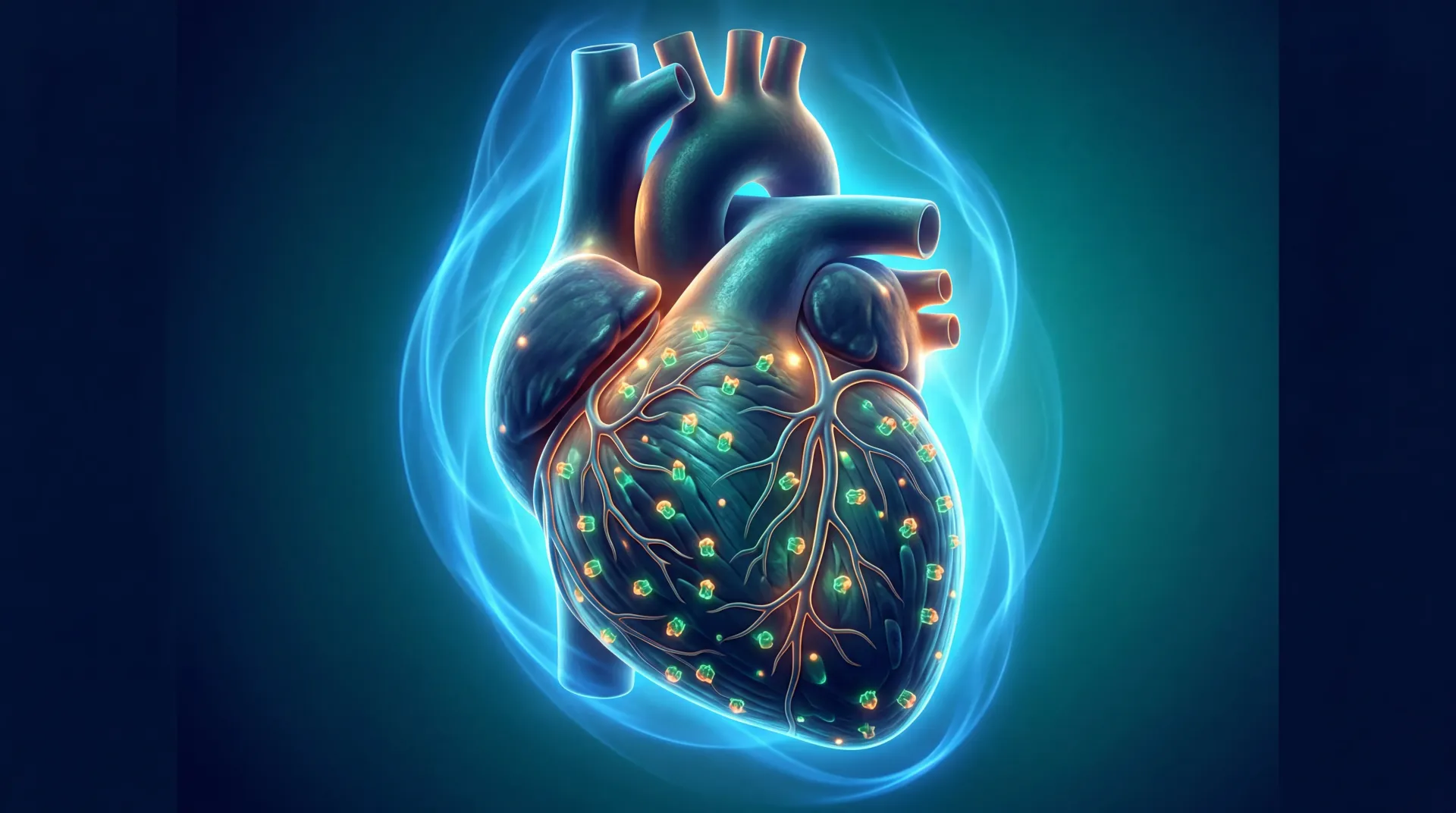
Sarcopenia (the age- or disease-related loss of muscle mass and function) is not a harmless consequence of getting older. When leg muscles atrophy, the body loses its metabolic buffer for glucose, inflammation rises, and the heart's workload steadily increases. Your thigh muscles are actively protecting your cardiovascular system every day.
Your Muscles Are a Pharmacy (If You Use Them)
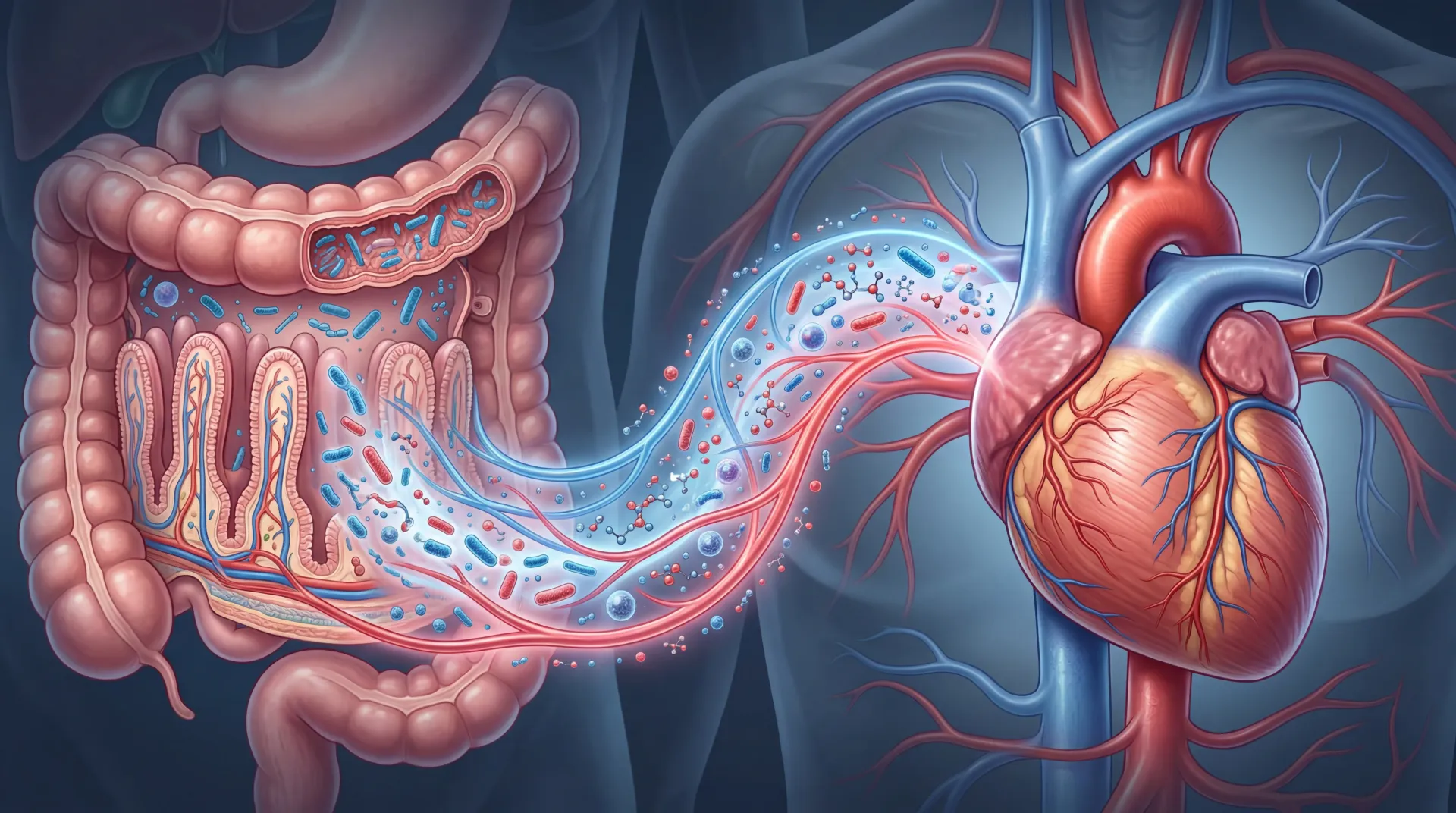
Skeletal muscle accounts for roughly 40% of body weight in non-obese adults. It is now recognized as the body's largest secretory organ. During intense contraction, especially against heavy resistance, muscle fibers release hundreds of signaling molecules called myokines. These peptides travel through the bloodstream to the liver, fat tissue, brain, gut, and heart, functioning as chemical messengers that regulate inflammation, blood sugar, and fat metabolism.
Two myokines matter most for heart protection:
Interleukin-6 (IL-6) has a split personality. When released by immune cells during infection, it drives inflammation. But muscle-derived IL-6, released during exercise, does the opposite. It directly blocks TNF-alpha (a major driver of atherosclerosis), stimulates anti-inflammatory cytokines IL-1ra and IL-10, increases glucose uptake into muscle cells through GLUT4 translocation via AMPK activation, triggers fat breakdown in adipose tissue, and even protects pancreatic beta-cells from stress-induced death. It also stimulates GLP-1 release from intestinal L-cells, improving insulin secretion after meals.
Irisin is a peptide cleaved from the FNDC5 protein during muscular contraction. Meta-analyses show that resistance exercise triggers a larger irisin response than endurance exercise. Irisin acts on white fat tissue to convert it into metabolically active brown-like fat through UCP1 upregulation, a process called "browning." This transformation increases resting energy expenditure and reduces insulin resistance. Irisin also crosses the blood-brain barrier to stimulate brain-derived neurotrophic factor (BDNF), and experimental models show it can reduce heart attack damage by activating protective autophagy pathways and neutralizing reactive oxygen species in blood vessel walls.
| Myokine | Trigger | Targets | Cardiovascular Benefit |
|---|---|---|---|
| IL-6 (muscle-derived) | Resistance and aerobic contraction | Liver, fat tissue, immune cells, pancreas | Blocks TNF-alpha; increases glucose uptake; drives fat breakdown; protects beta-cells |
| Irisin | Muscle contraction (especially resistance) | White fat, heart, brain | Fat browning via UCP1; reduces infarct size; stimulates BDNF |
| Myonectin (CTRP15) | Endurance and resistance exercise | Heart muscle, macrophages | Protects against ischemic injury via S1P/cAMP/Akt pathways |
| FSTL-1 | Exercise | Blood vessels, endothelium | Improves endothelial function; drives new blood vessel formation |
Other myokines include myonectin, which protects the heart from ischemic injury, and FSTL-1, which promotes blood vessel repair through nitric oxide pathways. The combined effect is a whole-body anti-inflammatory, anti-diabetic, anti-atherosclerotic chemical environment, but only when muscle is actually contracting under load.
When Weak Muscles Accelerate Heart Disease
While exercising muscles release protective molecules, failing muscles release destructive ones. Myostatin (growth differentiation factor 8), part of the TGF-beta superfamily, normally limits muscle growth to prevent unchecked hypertrophy. But in heart failure, both the failing heart and atrophying muscles ramp up myostatin production dramatically.
This creates a feedback loop. The failing heart releases myostatin that directly causes skeletal muscle wasting (cardiac cachexia). Genetic deletion studies proved this definitively: removing myostatin from heart muscle completely abolished the skeletal muscle wasting typical of heart failure. The failing heart was commanding the body's muscles to self-destruct.
Elevated myostatin activates Smad2/Smad3 pathways that shut down protein synthesis and accelerate muscle breakdown. It also drives cardiac fibrosis, stiffening heart tissue and impairing both filling and pumping.
Resistance training is the most effective physiological counter to this cascade. Heavy loading downregulates myostatin expression while upregulating decorin and follistatin, natural myostatin blockers. In the presence of follistatin, myostatin cannot connect to its receptor, halting muscle wasting. This is why structured resistance training is now considered a frontline intervention even for patients with existing heart failure.
The Calf Muscle Pump: Your Body's Second Heart
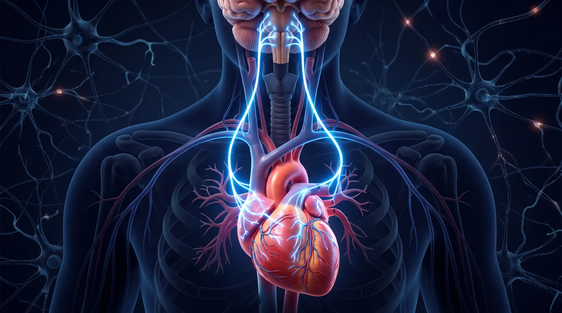
The relationship between leg muscles and heart health is not purely biochemical. It is mechanical. Your lower legs function as an auxiliary pump for blood circulation, often called the body's "second heart."
Gravity constantly works against venous return. Getting blood from your feet back to your heart is an engineering problem the heart alone cannot solve. The soleus and gastrocnemius muscles in the calf, working with deep venous valves, handle most of this work. During walking, running, squats, or standing exercises, calf contractions compress deep veins and propel blood upward. When the muscle relaxes, distal valves open to refill from the superficial system.
This compression-decompression cycle generates a pressure gradient that moves blood vertically and horizontally, equalizing pressure between deep and superficial veins. The result: increased venous return that optimizes cardiac preload and stroke volume via the Frank-Starling mechanism, reducing the heart's workload beat by beat.
When calf muscles are weak or inactive, blood pools in the lower legs, raising the risk of venous hypertension, edema, and deep vein thrombosis (DVT). The heart compensates by increasing its baseline workload, gradually exhausting its reserves. Strong calf muscles provide continuous mechanical offloading for the heart throughout your entire life.
How Heavy Lifting Cleans Your Arteries
Traditional cardiac rehab has focused on moderate aerobic exercise. But heavy resistance training creates vascular benefits that walking on a treadmill cannot replicate.
During heavy leg exercises like squats or leg presses, the massive intramuscular pressure briefly occludes arteries in the working limb. The moment the muscle relaxes, blood rushes back in (reactive hyperemia). This violent oscillation in flow creates high-magnitude shear stress against arterial walls, which is the primary mechanical trigger for endothelial nitric oxide synthase (eNOS) activation.
Activated eNOS produces nitric oxide (NO) from L-arginine. NO relaxes vascular smooth muscle, lowers peripheral resistance, and drops blood pressure. Consistent NO production also reverses arterial stiffness associated with aging and atherosclerosis. Clinical trials show that combined aerobic and resistance training increases basal NO production and decreases arterial stiffness (measured by pulse wave velocity) more effectively than either modality alone.
Aerobic exercise creates sustained, low-magnitude flow improvements. Resistance training creates intermittent, high-magnitude shear stress spikes. Combined training protocols produce synergistic endothelial health optimization that neither can achieve independently.
Muscles as Metabolic Sinks: The Diabetes Connection
Heart failure, coronary artery disease, and atherosclerosis are tightly linked to insulin resistance and type 2 diabetes. Skeletal muscle is the body's primary disposal system for blood sugar, responsible for clearing over 80% of postprandial glucose. When muscle mass declines, the body loses this metabolic buffer.
Heavy resistance training triggers mechanical translocation of GLUT4 glucose transporters to the muscle cell membrane. This happens through a pathway that bypasses insulin signaling entirely, which is exactly the mechanism that is broken in type 2 diabetes. Chronically, resistance training also increases total GLUT4 protein content, enhances mitochondrial function, and reduces toxic intramuscular fat accumulation.
People with severe insulin resistance typically have reduced capillary density around their muscle fibers and heavy immune cell infiltration that physically limits glucose transport. Targeted exercise training reverses both: new capillaries form, immune cells clear out, and lipid metabolites get burned, restoring glucose uptake and glycogen synthesis.
These improvements happen regardless of whether visible muscle size increases. What matters for metabolic health is the functional quality and cellular health of muscle fibers, not just their cross-sectional area. The repeated mechanical tension of resistance training embeds long-term metabolic adaptations into the tissue itself.
The 30-Second Chair Stand Test: A Life-or-Death Screening
Full cardiopulmonary exercise testing (VO2 peak measurement) requires expensive equipment, trained staff, and patients healthy enough to use a treadmill. Most frail or elderly patients cannot do it. The 30-Second Chair Stand Test (30sCST) has filled this gap as a validated proxy for both leg power and cardiorespiratory fitness.
The protocol: sit in a straight-backed chair (43.2 cm height), arms crossed over your chest, and stand fully upright then sit back down as many times as possible in 30 seconds. In heart failure patients (stage A/B), the number of stands correlates strongly with VO2 peak (AUC 0.740 for males, 0.725 for females).
Target cut-offs for adequate exercise tolerance are 18 stands for men and 16 for women. At the other end, patients managing fewer than 4-11 stands (depending on age and sex) face mortality hazard ratios over three times higher (HR ~3.31; 95% CI: 1.03-10.6) compared to those completing 11 or more. A poor performance signals that peripheral muscle weakness has crossed the threshold where skeletal muscle can no longer compensate for declining cardiac output.
| 30s Chair Stand Performance | Interpretation | Mortality Risk |
|---|---|---|
| 18+ stands (men) / 16+ (women) | Adequate exercise tolerance | Baseline |
| 11-17 stands | Moderate functional capacity | Elevated |
| Fewer than 4-11 stands (age-dependent) | Severe functional limitation | HR ~3.31 (3x higher) |
Current Guidelines: Squats Before Statins
The 2023 AHA Scientific Statement on Resistance Training permanently dismantled decades of clinical caution about lifting heavy weights with heart disease. Previous theories that resistance training would cause dangerous blood pressure spikes or tank ejection fraction in heart failure patients have been clinically disproven.
Resistance training is now prescribed as a first-line intervention for patients with heart failure (reduced ejection fraction), coronary artery disease, peripheral artery disease, and even end-stage kidney disease during dialysis. The measured benefits: resting systolic blood pressure drops by about 6 mmHg and diastolic by 5 mmHg in hypertensive individuals. These reductions rival first-line antihypertensive medications. Fasting glucose drops, HbA1c falls by up to 0.34%, HDL rises modestly, triglycerides and total cholesterol decrease, and type 2 diabetes incidence drops by 17%.
The 2025 ACC/AHA blood pressure guidelines formalized a "lifestyle first" sequence: for adults with elevated blood pressure (130/80 mmHg or higher) but below 7.5% 10-year cardiovascular risk, clinicians must prescribe 3-6 months of structured exercise (including resistance training), dietary changes (such as the DASH diet), and weight management before escalating to medications. Drugs come only after this trial period fails.
This prevents what the guidelines call the trap of chemically managing downstream symptoms (high blood pressure, high cholesterol) of what is fundamentally a muscular and mechanical deficit. Sarcopenia, absent shear stress, and lost glucose disposal capacity are root causes. Addressing them directly through structured exercise and dietary changes resolves the pathology at its source.
For acute events, the 2025 ACC/AHA acute coronary syndrome guidelines still mandate aggressive pharmacotherapy: dual antiplatelet therapy, high-intensity statins, and PCSK9 inhibitors if LDL-C stays above 70 mg/dL. But these are now paired with mandatory cardiac rehabilitation referrals, including home-based options with telehealth monitoring for the 80%+ of patients who skip in-person rehab.
Pre-habilitation is gaining ground too. Systematic reviews show that 4-12 weeks of pre-surgical exercise improves 6-Minute Walk Test distance by over 52 meters and shortens ICU stay by an average of 15 hours. The old rule of waiting 6-8 weeks after sternotomy for resistance work has been overturned: kinematic studies show early resistance training (within 4 weeks post-surgery) poses no greater sternal risk than coughing.
Frequently Asked Questions
How strong do my quadriceps need to be to protect my heart?
Based on the Yamamoto et al. study, the median thresholds that divided high from low strength were 33% of body weight for women and 52% for men. Each 5% increase in body-weight-relative quadriceps strength reduced heart failure risk by 11%. There is no ceiling to the benefit; stronger is consistently better.
Can resistance training replace my heart medications?
Not for acute events or high-risk profiles. Current guidelines use a sequenced approach: for adults with elevated blood pressure but lower overall cardiovascular risk, 3-6 months of structured exercise (including resistance training) is tried before adding medications. For people already on medications after a heart attack or with advanced disease, resistance training is added alongside pharmacotherapy, not instead of it. Always work with your cardiologist.
Is heavy lifting safe if I already have heart failure?
Yes, according to the 2023 AHA Scientific Statement. Fears that resistance training causes dangerous pressure spikes or worsens ejection fraction have been disproven. RT is now prescribed for heart failure, coronary artery disease, and peripheral artery disease patients. Start with supervised sessions and progress gradually.
What is the 30-Second Chair Stand Test and should I try it?
It is a validated screening tool where you sit in a standard chair, cross your arms, and stand up and sit down as many times as possible in 30 seconds. Target scores are 18+ stands for men and 16+ for women. Scoring below age-specific thresholds (as few as 4-11 stands) is associated with over three times higher mortality risk. It can be done at home as a rough self-assessment, but discuss results with your doctor.
How does the calf muscle pump actually help my heart?
Your calf muscles (soleus and gastrocnemius) compress deep veins during walking, running, or squatting, propelling blood upward against gravity back toward the heart. This increases venous return, optimizes the heart's stroke volume via the Frank-Starling mechanism, and reduces the heart rate and contractility demands on the heart. Weak or inactive calves force the heart to compensate by working harder to maintain circulation.
Related Articles
- Physical Exercise and Brain Health - How the same exercises that protect your heart also improve cognitive function and reduce neurodegeneration risk.
- Fitness, Pain Management, and Wellness Guide - A comprehensive look at structured exercise for pain reduction and overall wellness, including resistance training protocols.
- Diets That Prevent and Help Diabetes - Dietary strategies that complement resistance training for blood sugar control and reduced cardiovascular risk.
- Boost Your Metabolism Daily in 17 Easy Steps - Practical daily habits to increase metabolic rate, including the role of muscle mass in calorie expenditure.
- Exercises for Lower Back Pain Relief - Safe strengthening exercises that complement a leg training program while addressing common back pain.
Medical Disclaimer
This article is for informational and educational purposes only and is not medical advice, diagnosis, or treatment. Always consult a licensed physician or qualified healthcare professional regarding any medical concerns. Never ignore professional medical advice or delay seeking care because of something you read on this site. If you think you have a medical emergency, call 911 immediately.